Fascial Layers, Part 2 + Anatomy of a Nerve

For the most part, fascia can be classified as either superficial or deep, with the superficial layer being just beneath the skin and the deep layer being, well, everything else. As far as the deep layer goes, the “everything else” can be classified as either meningeal fascia or visceral fascia. 

DEEP FASCIA

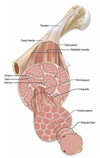
Joint capsules, ligaments, tendons and the three layers that weave their way through each muscle (epimysium, perimysium and endomysium) make up the deep fascia. Aponeuroses are included in this classification as well. An aponeurosis is a broad, thick sheet of connective tissue that serves as an attachment site for muscles. A big one is the diamond-shaped thoracolumbar aponeurosis (where the latissimus dorsi originates) that spans across the lower back. I explained in Part 1 that fascia is rich in lots of sensory receptors. I’ll go into detail about each of those in a later post, but one of the sensory receptors found in fascia is called a nociceptor, otherwise known as a pain receptor. I once came across a study showing that people with lower back pain had a thickening of the thoracolumbar fascia compared to those in the trial who didn’t suffer from any LBP. This makes sense because with pain receptors being spread all throughout the fascial layers, a thickening of the fascia would equal an increase in pain receptors.

VISCERAL FASCIA

The visceral fascia is the layer that surrounds the heart (specifically called the pericardium) and lungs (pleura) as well as the abdominal organs. It also suspends the organs within their respective cavities (thoracic or abdominal) by way of ligaments that are meant to hold the organs against the body wall as well as allow for necessary physiological movement like breathing, the heart beating and peristalsis. 

MENINGEAL FASCIA

This is the layer that surrounds the brain and the nervous system. To better understand this, we’ll look at the anatomy of a nerve. But first...

WHAT IS A NERVE?

A nerve is a structured pathway that allows for the transmission of impulses to and from the brain and nervous system. Nerves either have one type of neuron, in which case they are classified as sensory or motor nerves, or like most nerves they have both motor and sensory neurons and are called mixed nerves. Nerves are structurally very similar to skeletal muscle in that each nerve has three separate layers of fascia, just like each muscle. 

Let’s look at the structure of a nerve from superficial to deep. The outer fascial covering of a nerve is called the epineurium (translates to on the nerve). Inside of that, nerve fibers (also called axons) are bundled together the same way muscle fibers are bundled, the layer that surrounds each bundle of axons is called the perineurium (around the nerve). Each individual axon that makes up the bundle is also surrounded by its own layer of fascia, this is called the endoneurium (within the nerve). 

Unknown-1.jpeg

Quick Trapezius Overview

The trapezius muscle is huge and has fibers that run in multiple directions which gives it the ability to do lots of different things. As a whole, the trapezius originates at the middle portion of the base of the skull (external occipital protuberance and the medial portion of the superior nuchal line or you can just say middle of base of skull), it latches on to the big thick neck ligament and then grabs on to the spinous processes of C7-T12. It inserts itself on the lateral third of the clavicle, the acromion and the spine of the scapula. 

Probably everyone knows where their upper traps are because nearly everyone complains about how tight they are. What tends to happen is the upper traps get overdeveloped while the middle and lower traps get weak. Learning how to use the middle and lower traps to retract and depress the scapulae rather than trying to force them down with the the upper traps (which don’t have the pulling capacity to do that at all) can be helpful in reducing shoulder and neck tension/pain and headaches. 

The upper fibers of the trapezius muscle are very involved in head and neck movement. The traps laterally flex the head and neck, rotate (turn) the head to the opposite side (so if you look to the right, your left upper trap is contracting) and when both the left and right upper trapezius muscles fire together, they extend the head and neck. In addition to that they also elevate the scapula and they upwardly rotate it as well. 

Moving down to the middle fibers... it makes sense that they retract and stabilize the scapula because the origin is on the spine. If the middle fibers of the trapezius which are perpendicular to the spine contract, the origin will pull the insertion toward it which draws the shoulder blades in toward the spine. Think about someone placing their fingers between your shoulder blades and then you gently pinching them by drawing the shoulder blades together. This activates the middle fibers of trapezius.

If you look at the fibers of the lower trapezius you can see that they run in a more downward direction. This gives them the leverage they need to depress the scapula or draw the shoulder blades down the back. 

Try this: Place your right hand on your left upper trap and see how it feels. Then relax your arms and shake them off a little bit. From there, gently gather the shoulder blades together, and then imagine sliding just the bottom tips of the shoulder blades down and into your back pockets. Maintain that and take the right hand to the left upper trap again and see how it feels softer with the support coming from the middle and lower fibers below.

Unknown.png

Fascial Layers, Part 1 + Anatomy of a Muscle

While it’s true that the fascia is one big continuous and completely connected piece of tissue, it looks and acts differently depending on where it’s located in the body. In order to gain a better understanding of the fascial system as a whole and also have it be less overwhelming, we’ll break it down into more digestible bites. 

Just beneath your skin there is a layer of fatty tissue that provides the body with necessary insulation, blood and lymphatic flow and energy storage. Just beneath that is the superficial fascia. It anchors the skin to the tissues and organs below and is rich in blood and lymphatic vessels, nerves and some general sensory receptors which I’ll describe in detail in a later post. This thin and fibrous but highly elastic layer is classified as loose connective tissue. In this case loose just means it lacks any regular pattern or strong organization. 

Unlike the superficial fascia, the deep fascia is dense and well-organized. As far as I’m concerned, it’s the coolest layer of fascia because of the incredible and stunningly beautiful way it surrounds, supports and separates yet also connects every single structure in your body. The deep fascia is rich in sensory receptors that are sensitive to things like pressure and movement, which I will also cover in detail in another post. First let’s look at the anatomy of a muscle. Understanding the fascial anatomy of a muscle is essential for truly understanding how yoga and massage create change for people and actually really “work”.

Let’s consider a muscle from the outside in, or anatomically speaking, superficial to deep. Every muscle as a whole is wrapped in a sleeve of fascia called the epimysium. Epi- meaning on and my- meaning muscle. Epimysium means on the muscle.

Within each muscle are groups of muscle cells that have been bundled together into what’s called fascicles. Each fascicle is wrapped in its own layer of fascia called the perimysium. Peri- meaning around and my- meaning muscle. Perimysium means around the muscle. 

Each individual muscle cell also has its own layer of fascia called the endomysium. Endo- meaning within, my- meaning muscle. Endomysium meaning within the muscle. 

Each of these three layers comes together to form the tendons that connect muscle to bone. The layer of fascia that surrounds each bone is called the periosteum.

In part two we’ll look at the fascial layers that surround the brain, nerves and organs as well as the anatomy of a nerve.

Unknown.jpeg