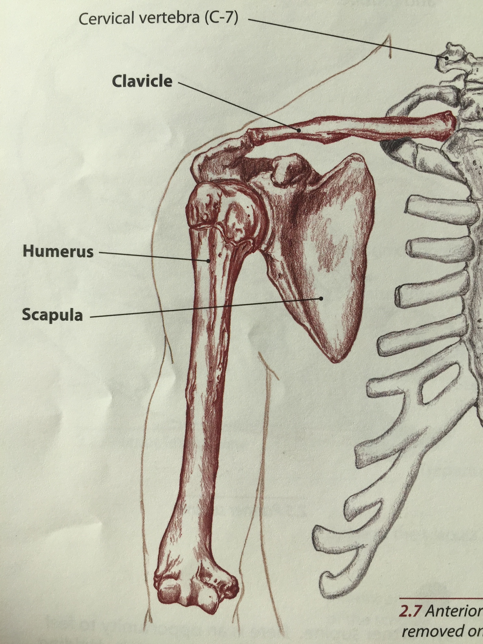
In part one we looked at the bony anatomy of the scapula, the first of three bones that make up the shoulder. In part 2 we will look at the clavicle and humerus.
Let’s start with the clavicle, or collar bone. The clavicle is the first bone to start ossifying (hardening) in the human fetus at about 6 weeks. Interestingly enough it is also the last to fully develop, this doesn’t usually happen until the late teens or early twenties. This combined with the fact that the clavicle is quite superficial makes it one of the most frequently broken bones in the body. When we’re talking about the clavicle it’s broken down into thirds: the lateral third (articulates with the acromion, also called the acromial end), the middle third and the medial third (articulates with the sternum, called the sternal end). The clavicle is pretty simple, it does have a few landmarks but I won’t mention them here.

A view of the clavicle from above.
The humerus has a bit more going on than the clavicle, and for our purposes we’ll just look at the proximal half for now (the half that’s closest to the shoulder). A review of scapular anatomy might be helpful, you can find that here.

A view of the right humerus from the front.
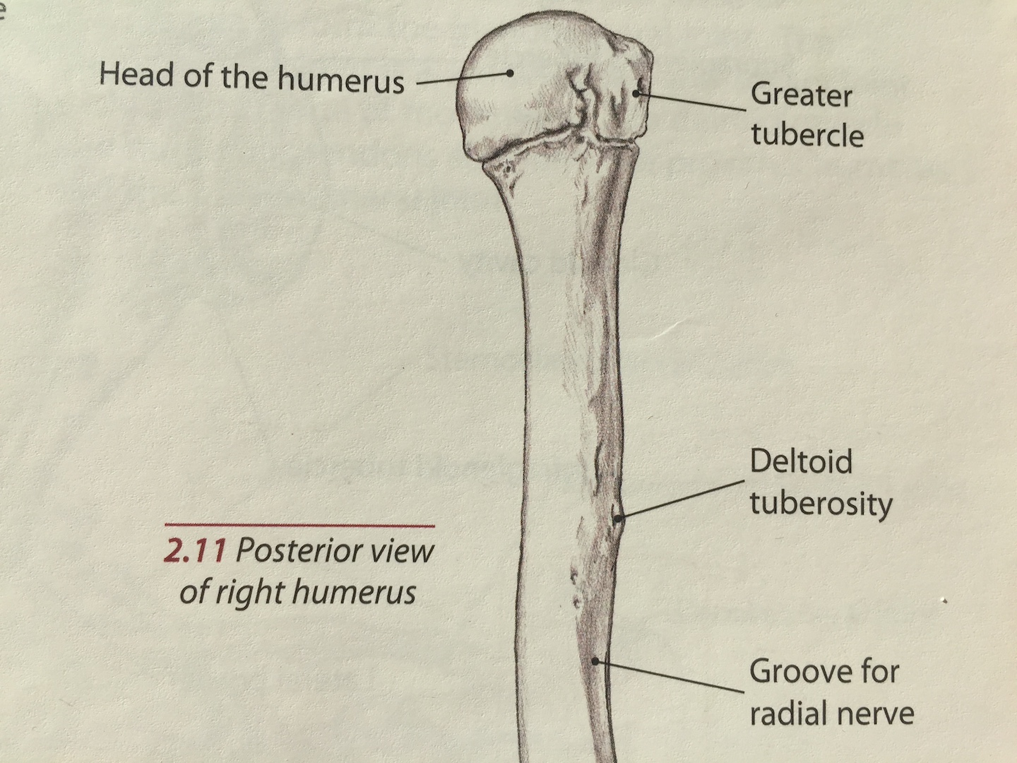
A view of the right humerus from the back.
THE HUMERUS
Head of the humerus - the head of the humerus sits in the glenoid fossa like a golf ball on a tee. The head of the humerus is the golf ball, the shallow glenoid fossa is the tee. This creates the glenohumeral joint, which we will get to in another post that covers joints and other structures.
Greater tubercle - this is the attachment site for three of the four rotator cuff muscles. It’s a bony knob on the humeral head that sits a little bit posterior (behind) and lateral (away from the midline) to its counterpart, the lesser tubercle.
Lesser Tubercle - a smaller bony knob that sits just medial (closer to the midline) and anterior (in front of) to the greater tubercle. This is the insertion point for the only muscle of the rotator cuff that doesn’t insert on the greater tubercle.
Crest of greater tubercle - Crest is a Latin term meaning ridge. It’s just distal to the greater tubercle.
Crest of the lesser tubercle - The ridge that comes down from the lesser tubercle, sits medial to the intertubercular groove.
Intertubercular groove - this lives in between the crests of the greater and lesser tubercles and is also called the bicipital groove, as the biceps brachii muscle feeds through here. You can imagine the crests as the peaks and the intertubercular groove as the valley. Intertubercular means in between the grooves. A lot of times a part of the body will tell you where it is, how big or small it is, what it does or some combination of the three based on its name.
Deltoid tuberosity - this is as far down the humerus as we’ll go for now. The deltoid tuberosity is found about halfway down the humerus, it’s a little bony knob where the all the fibers of the deltoid insert. It’s on the lateral humerus (outside).
Familiarize yourself with this information and it will be easier to understand the muscles and what they do (and how) once we start talking about them.